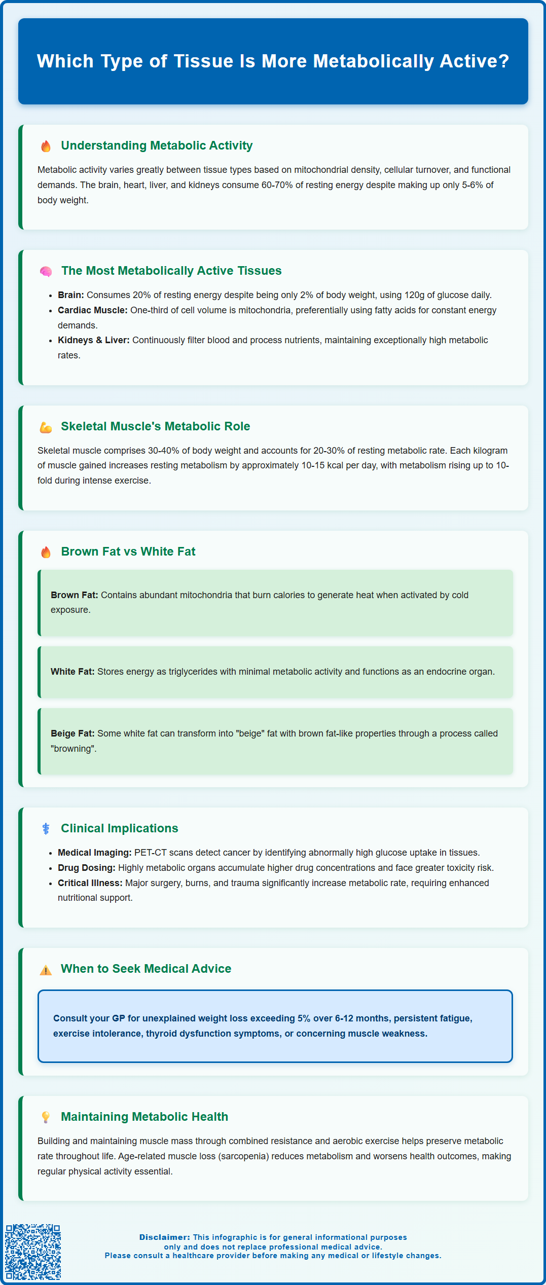
Which type of tissue is more metabolically active? Understanding metabolic activity across different human tissues is fundamental to comprehending energy expenditure, physiological function, and clinical disease. Metabolic activity refers to the rate at which cells consume energy and oxygen to maintain their functions. Tissues vary dramatically in their metabolic rates, determined by mitochondrial density, cellular turnover, and functional demands. Highly metabolic tissues—including the brain, heart, kidneys, and liver—account for approximately 60–70% of total body energy expenditure despite representing only 5–6% of body weight. This knowledge underpins diagnostic imaging, nutritional management, pharmacology, and weight management strategies across clinical practice.
Summary: Cardiac muscle, kidneys, and brain tissue are amongst the most metabolically active tissues per gram, with the brain consuming approximately 20% of resting metabolic rate despite being only 2% of body weight.
- Metabolic activity is determined by mitochondrial density, cellular turnover rate, functional demands, and tissue vascularisation.
- The brain, heart, liver, and kidneys account for 60–70% of total body energy expenditure despite representing only 5–6% of body weight.
- Brown adipose tissue can achieve very high metabolic rates when activated by cold exposure, unlike white adipose tissue which primarily stores energy.
- Skeletal muscle contributes 20–30% of resting metabolic rate and increases dramatically during physical activity, making it a key target for metabolic health interventions.
- PET-CT scanning exploits tissue metabolic differences using radiolabelled glucose to detect malignancies and assess physiologically active tissues.
- Patients should seek GP advice for unexplained weight loss, persistent fatigue, suspected thyroid dysfunction, or concerns about muscle weakness.
Table of Contents
Understanding Metabolic Activity in Human Tissues
Metabolic activity refers to the rate at which cells within tissues consume energy and oxygen to maintain their functions. Different tissue types exhibit vastly different metabolic rates, determined by their cellular composition, mitochondrial density, and physiological roles. Understanding these differences is fundamental to comprehending human physiology, energy expenditure, and various clinical conditions.
Key factors influencing tissue metabolic activity include:
-
Mitochondrial density – tissues with more mitochondria generate more ATP and consume more oxygen
-
Cellular turnover rate – rapidly dividing or regenerating tissues require substantial energy
-
Functional demands – organs performing continuous work (such as the heart) maintain high metabolic rates
-
Vascularisation – well-perfused tissues typically show enhanced metabolic activity
Metabolic rate is commonly measured using oxygen consumption (VO₂), glucose uptake via PET scanning, or heat production through calorimetry. In healthy adults at rest under thermoneutral conditions, the brain, heart, liver, and kidneys account for approximately 60-70% of total body energy expenditure despite representing only about 5-6% of body weight. This disproportionate energy consumption reflects their essential, continuous physiological functions, though these proportions vary with age, body composition and environmental conditions.
Clinically, understanding tissue metabolic activity has important implications for nutritional requirements, drug distribution, imaging techniques, and disease processes. Conditions affecting highly metabolic tissues often present acutely and require prompt intervention. Furthermore, metabolic activity influences wound healing, immune responses, and the body's adaptation to physiological stress. The concept also underpins modern approaches to weight management, as tissues with higher metabolic rates contribute more substantially to total daily energy expenditure.

Which Type of Tissue Is More Metabolically Active?
Among the body's tissues, several demonstrate exceptionally high metabolic activity, with different tissues showing prominence depending on whether they are at rest or in an activated state.
When comparing major tissue categories by metabolic rate per gram of tissue, the hierarchy typically follows this pattern:
-
Cardiac muscle – continuously contracting tissue with dense mitochondrial content and high sustained metabolic rate
-
Kidneys – high energy demands due to constant filtration and reabsorption processes
-
Brain tissue – accounts for approximately 20% of resting metabolic rate despite being only 2% of body weight
-
Liver – central metabolic organ performing hundreds of biochemical reactions simultaneously
-
Brown adipose tissue (BAT) – variable but potentially high metabolic rate, particularly when activated by cold exposure
-
Skeletal muscle – moderate resting metabolic rate; significant contributor due to total mass
-
White adipose tissue – relatively low metabolic activity, primarily serving energy storage
The brain's metabolic activity remains remarkably constant, consuming approximately 120g of glucose daily in adults under normal conditions, though it can utilise ketones during prolonged fasting. This continuous high demand makes the brain particularly vulnerable to hypoglycaemia and hypoxia. Cardiac muscle similarly maintains constant activity, with metabolic rate increasing proportionally during physical exertion or stress.
Skeletal muscle presents an interesting case: whilst individual muscle fibres have moderate metabolic rates at rest, the sheer volume of skeletal muscle in the body (typically 30-40% of body weight) means it contributes substantially to total energy expenditure. During intense exercise, local muscle metabolism can increase many-fold, while whole-body oxygen consumption typically rises up to 10-fold at maximal exertion.
Brown adipose tissue, when maximally activated by cold exposure or sympathetic stimulation, can achieve very high metabolic rates per gram, though its total contribution to whole-body energy expenditure in adult humans is typically modest due to its limited mass.
Brown Fat vs White Fat: Metabolic Differences
The distinction between brown adipose tissue (BAT) and white adipose tissue (WAT) represents one of the most striking examples of metabolic diversity within a single tissue type. These two forms of adipose tissue serve fundamentally different physiological purposes and exhibit vastly different metabolic profiles.
Brown adipose tissue characteristics:
-
Contains numerous small lipid droplets (multilocular) rather than one large droplet
-
Extremely high mitochondrial density, giving the tissue its characteristic brown colour
-
Expresses UCP1, which dissipates the proton gradient to generate heat rather than ATP
-
Highly vascularised and innervated by sympathetic nerves
-
Activated by cold exposure and certain hormones (noradrenaline, thyroid hormones)
-
More prevalent in infants; adults retain deposits in the neck, supraclavicular regions, and along the spine
When activated, brown fat can significantly increase its metabolic rate, consuming substantial amounts of glucose and fatty acids whilst generating heat. PET-CT imaging studies have demonstrated that active BAT can contribute to glucose disposal, leading to research interest in its potential role in metabolic health.
White adipose tissue characteristics:
-
Contains a single large lipid droplet (unilocular) occupying most of the cell
-
Relatively few mitochondria and low baseline metabolic activity
-
Primary function is energy storage as triglycerides
-
Also serves endocrine functions, secreting adipokines including leptin and adiponectin
-
Distributed subcutaneously and viscerally throughout the body
Recent research has identified "beige" or "brite" adipocytes within white fat depots that can acquire brown fat-like characteristics under certain conditions, a process termed "browning". This phenomenon has generated considerable scientific interest regarding potential therapeutic applications for obesity and metabolic disorders, though clinical applications remain investigational. There are currently no MHRA-licensed medicines to induce 'browning' for weight management, and no official link has been established between pharmacological browning interventions and improved long-term metabolic outcomes in humans.
Muscle Tissue and Metabolic Rate
Muscle tissue, comprising both skeletal and cardiac varieties, plays a crucial role in whole-body metabolic rate, though the two types exhibit distinct metabolic profiles. Understanding muscle metabolism is essential for managing conditions ranging from sarcopenia to metabolic syndrome.
Skeletal muscle metabolism:
Skeletal muscle accounts for approximately 30-40% of body weight in healthy adults and contributes roughly 20-30% of resting metabolic rate. This contribution increases dramatically during physical activity. Muscle tissue is metabolically heterogeneous, containing different fibre types with varying metabolic characteristics:
-
Type I fibres (slow-twitch) – high mitochondrial content, oxidative metabolism, fatigue-resistant, used for endurance activities
-
Type II fibres (fast-twitch) – lower mitochondrial density, rely more on glycolytic metabolism, generate force rapidly but fatigue quickly
The metabolic activity of skeletal muscle is highly adaptable. Regular resistance training increases muscle mass and can elevate resting metabolic rate by approximately 10-15 kcal per day for each kilogram of muscle gained. Endurance training enhances mitochondrial density and oxidative capacity without necessarily increasing muscle size. This adaptability makes skeletal muscle a key target for interventions aimed at improving metabolic health.
Cardiac muscle metabolism:
The heart is an obligate aerobic organ with exceptionally high and constant metabolic demands. Cardiac muscle contains a very high mitochondrial density (approximately one-third of cell volume) and exhibits minimal capacity for anaerobic metabolism. The heart preferentially metabolises fatty acids (60-70% of energy) under normal conditions but can switch to glucose, lactate, or ketones depending on substrate availability and physiological state.
Clinically, maintaining adequate muscle mass is increasingly recognised as important for metabolic health. NICE guidance on obesity management (CG189) emphasises the role of physical activity in preserving muscle tissue during weight loss. The UK Chief Medical Officers' Physical Activity Guidelines recommend both resistance and aerobic exercise for health benefits. Sarcopenia (age-related muscle loss) is associated with reduced metabolic rate, increased frailty, and poorer health outcomes. Patients should be encouraged to engage in both resistance and aerobic exercise to maintain muscle mass and metabolic function throughout life.
Clinical Implications of Tissue Metabolic Activity
Understanding tissue metabolic activity has numerous practical applications in clinical medicine, influencing diagnostic approaches, treatment strategies, and patient management across multiple specialties.
Diagnostic imaging applications:
PET-CT scanning exploits differences in tissue metabolic activity by using radiolabelled glucose (¹⁸F-FDG). Malignant tissues typically exhibit markedly elevated glucose uptake compared to surrounding normal tissue, making PET-CT valuable for cancer detection, staging, and monitoring treatment response. However, physiologically active tissues (brain, heart, brown fat) also demonstrate high tracer uptake, which must be distinguished from pathological processes. NHS preparation guidelines typically advise patients to fast for approximately 6 hours before the scan, avoid strenuous exercise for 24 hours, and keep warm before the procedure to minimise brown fat activation that could complicate image interpretation.
Pharmacological considerations:
Drug distribution and metabolism are influenced by tissue metabolic activity. Highly perfused, metabolically active organs (liver, kidneys, brain) typically achieve higher drug concentrations and may be more susceptible to drug-related toxicity. The MHRA provides guidance on monitoring for organ-specific adverse effects with various medications, particularly those metabolised hepatically or cleared renally. Patients should report suspected side effects via the MHRA Yellow Card scheme (yellowcard.mhra.gov.uk).
Nutritional and metabolic management:
Patients with conditions affecting highly metabolic tissues often have increased nutritional requirements. Critical illness, major surgery, burns, and trauma substantially elevate metabolic rate, necessitating careful nutritional support. NICE guidance on nutrition support in adults (CG32) emphasises the importance of assessing metabolic demands and providing adequate calories and protein to prevent catabolism.
Weight management implications:
The relationship between tissue composition and metabolic rate informs weight management strategies. Preserving or increasing muscle mass during weight loss helps maintain metabolic rate and improves long-term outcomes. NICE guidance (CG189) recommends combined dietary modification and physical activity for sustainable weight management.
When to seek medical advice:
Patients should contact their GP if they experience:
-
Unexplained weight loss (particularly >5% of body weight over 6-12 months)
-
Persistent fatigue or exercise intolerance
-
Symptoms suggesting thyroid dysfunction (which profoundly affects tissue metabolic rate)
-
Concerns about muscle weakness or loss
Understanding tissue metabolic activity also informs rehabilitation strategies, wound healing management, and the physiological response to stress or illness. Healthcare professionals should consider metabolic demands when planning interventions and setting realistic recovery expectations for patients.
Frequently Asked Questions
Which tissue has the highest metabolic rate in the body?
Cardiac muscle, kidneys, and brain tissue exhibit the highest metabolic rates per gram of tissue. The brain alone consumes approximately 20% of the body's resting energy expenditure despite representing only 2% of body weight, whilst cardiac muscle maintains constant high metabolic activity due to continuous contraction.
How does brown fat differ from white fat metabolically?
Brown adipose tissue contains numerous mitochondria and expresses UCP1 protein, enabling it to generate heat and achieve very high metabolic rates when activated by cold exposure. White adipose tissue has few mitochondria, low baseline metabolic activity, and primarily functions for energy storage rather than heat production.
Does muscle tissue contribute significantly to metabolic rate?
Yes, skeletal muscle accounts for 30–40% of body weight and contributes approximately 20–30% of resting metabolic rate in healthy adults. This contribution increases substantially during physical activity, and maintaining muscle mass through resistance and aerobic exercise is important for metabolic health throughout life.
The health-related content published on this site is based on credible scientific sources and is periodically reviewed to ensure accuracy and relevance. Although we aim to reflect the most current medical knowledge, the material is meant for general education and awareness only.
The information on this site is not a substitute for professional medical advice. For any health concerns, please speak with a qualified medical professional. By using this information, you acknowledge responsibility for any decisions made and understand we are not liable for any consequences that may result.
Heading 1
Heading 2
Heading 3
Heading 4
Heading 5
Heading 6
Lorem ipsum dolor sit amet, consectetur adipiscing elit, sed do eiusmod tempor incididunt ut labore et dolore magna aliqua. Ut enim ad minim veniam, quis nostrud exercitation ullamco laboris nisi ut aliquip ex ea commodo consequat. Duis aute irure dolor in reprehenderit in voluptate velit esse cillum dolore eu fugiat nulla pariatur.
Block quote
Ordered list
- Item 1
- Item 2
- Item 3
Unordered list
- Item A
- Item B
- Item C
Bold text
Emphasis
Superscript
Subscript


